Definition
Neuropathic arthropathy
- progressive destructive arthropathy 2° to neurological condition
- usually minimal to no trauma
Etiology
Diabetes
Leprosy / syphilis
Other - polio / paraplegia / syringomyelia
Pathophysiology
1. Neuro-traumatic theory - cumulative trauma in insensate foot
2. Neurovascular theory
- neurally stimulated vascular reflex stimulates bone resorption
Eichenholtz Classification
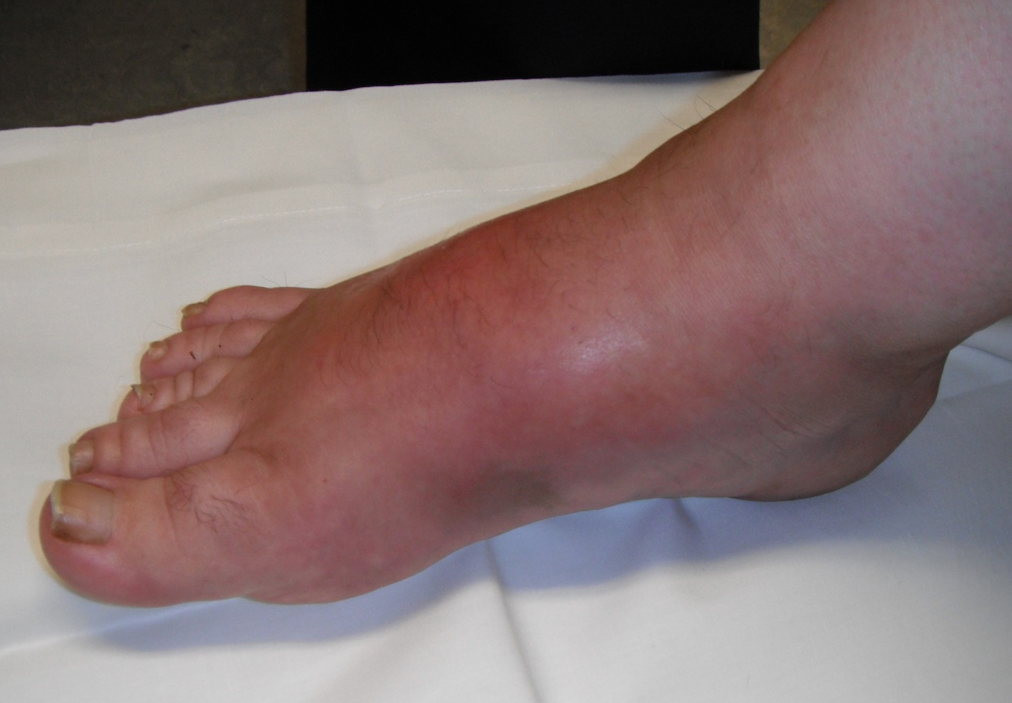
Stage 0
- added by Shibata et al 1990
- clinical signs (swelling/ erythema) precede XRay changes
- NWB during this period may prevent XRay changes
| Stage 1 Dissolution | Stage 2 Coalescence | Stage 3 Reconstruction | |
|---|---|---|---|
| Findings |
Acute inflammation (swollen, red, warm) Erythema reduces with elevation 10 minutes |
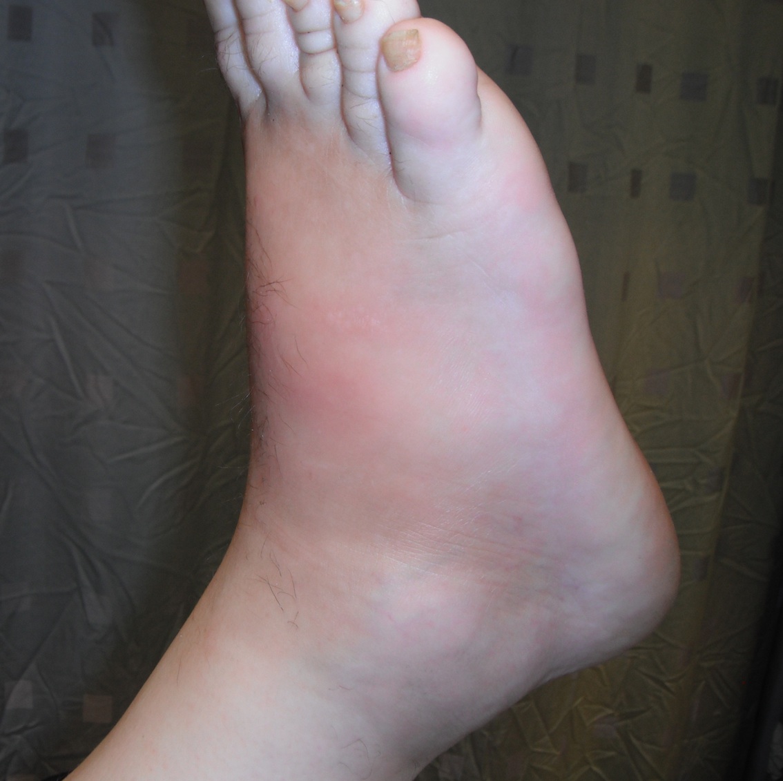
Inflammation decreases Reduced swelling Reduced temperature
|
Normal temperature Swelling reduced |
| Xray |
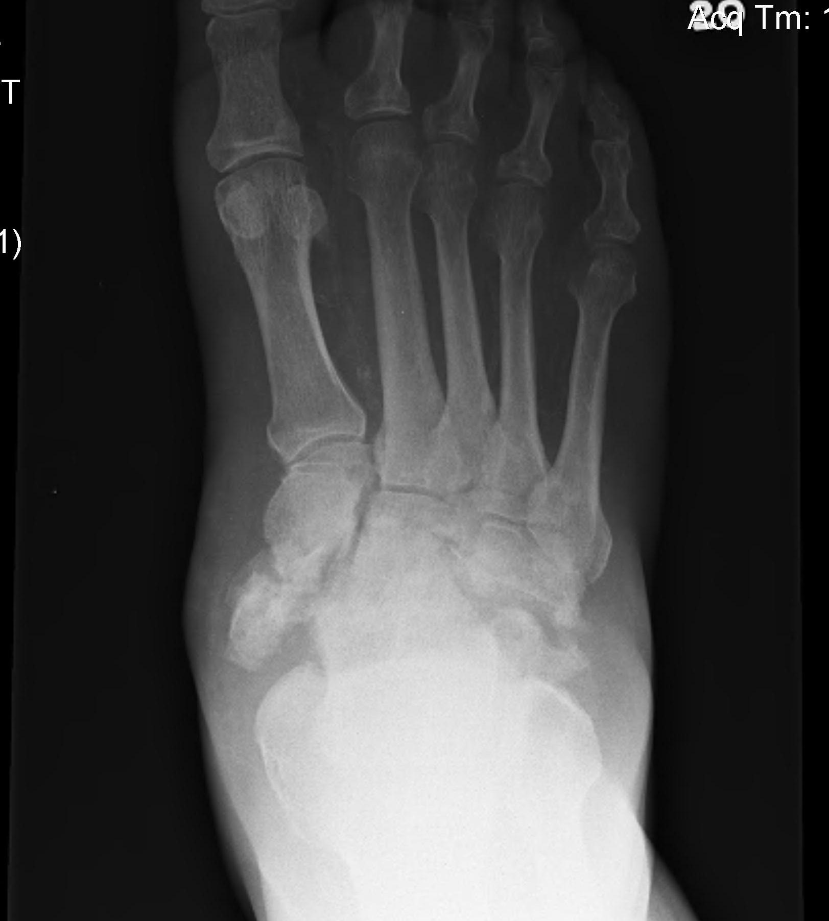
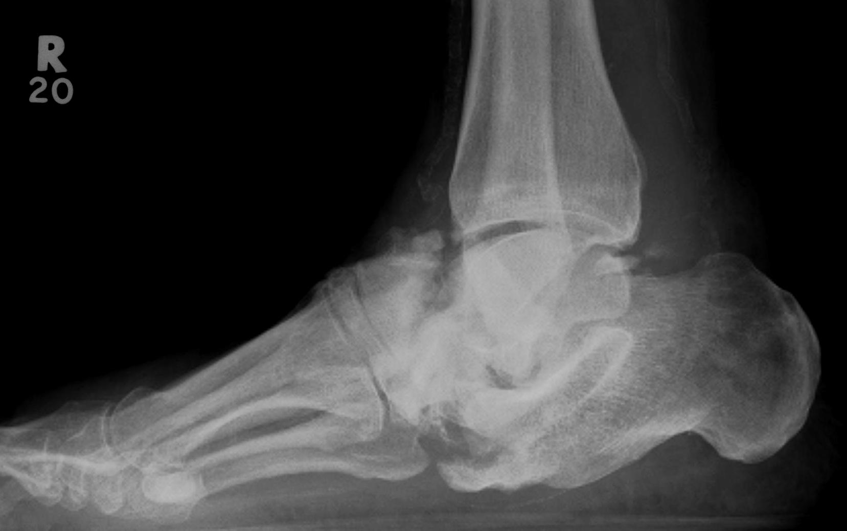
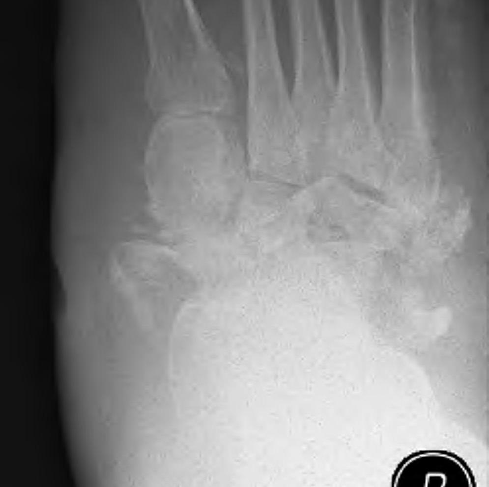
Demineralisation of regional bone Periarticular fragmentation Joint dislocation |
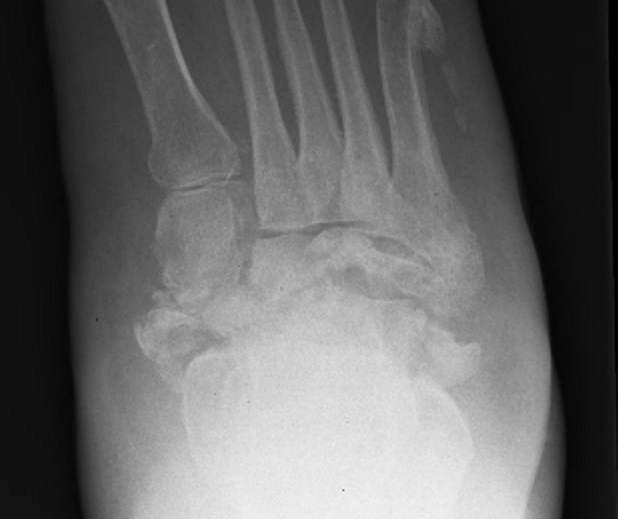
Absorption of osseous debris Organization and early healing of fracture fragments Periosteal new bone formation |
Smoothing of edges Sclerosis, osseous or fibrous ankylosis Bone healing Resolution of osteopenia
|
| Management |
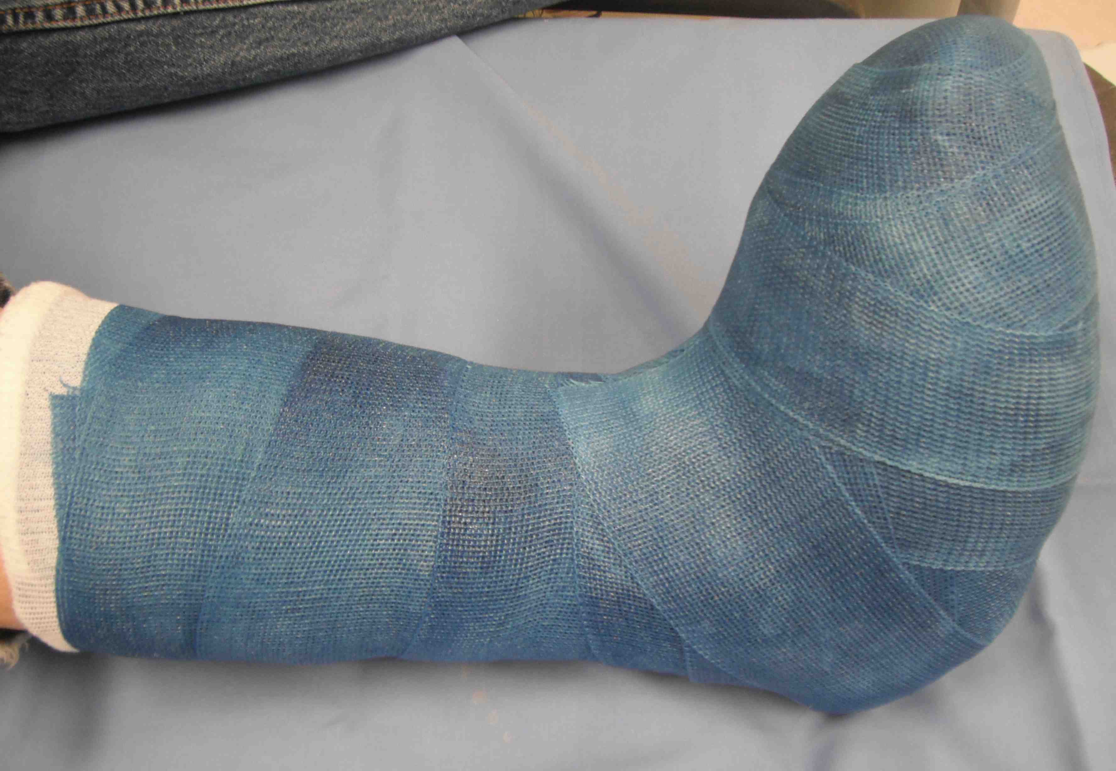
Total contact cast until stage 2 FWB |
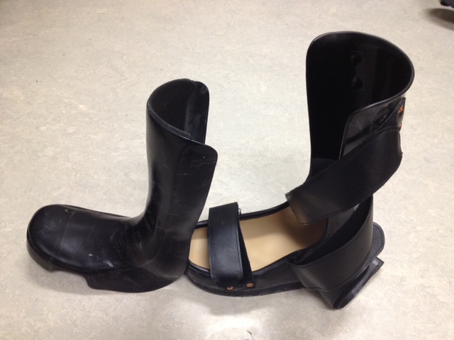
CROW (Charcot Resistant Orthotic Walker) Bivalved AFO |
Accommodative shoes with custom moulded orthotic
CROW or AFO if ongoing ankle instability |
 |
 |
|
Natural history
30% will relapse between stages
7% risk of BKA without ulcer
28% risk of BKA with ulceration
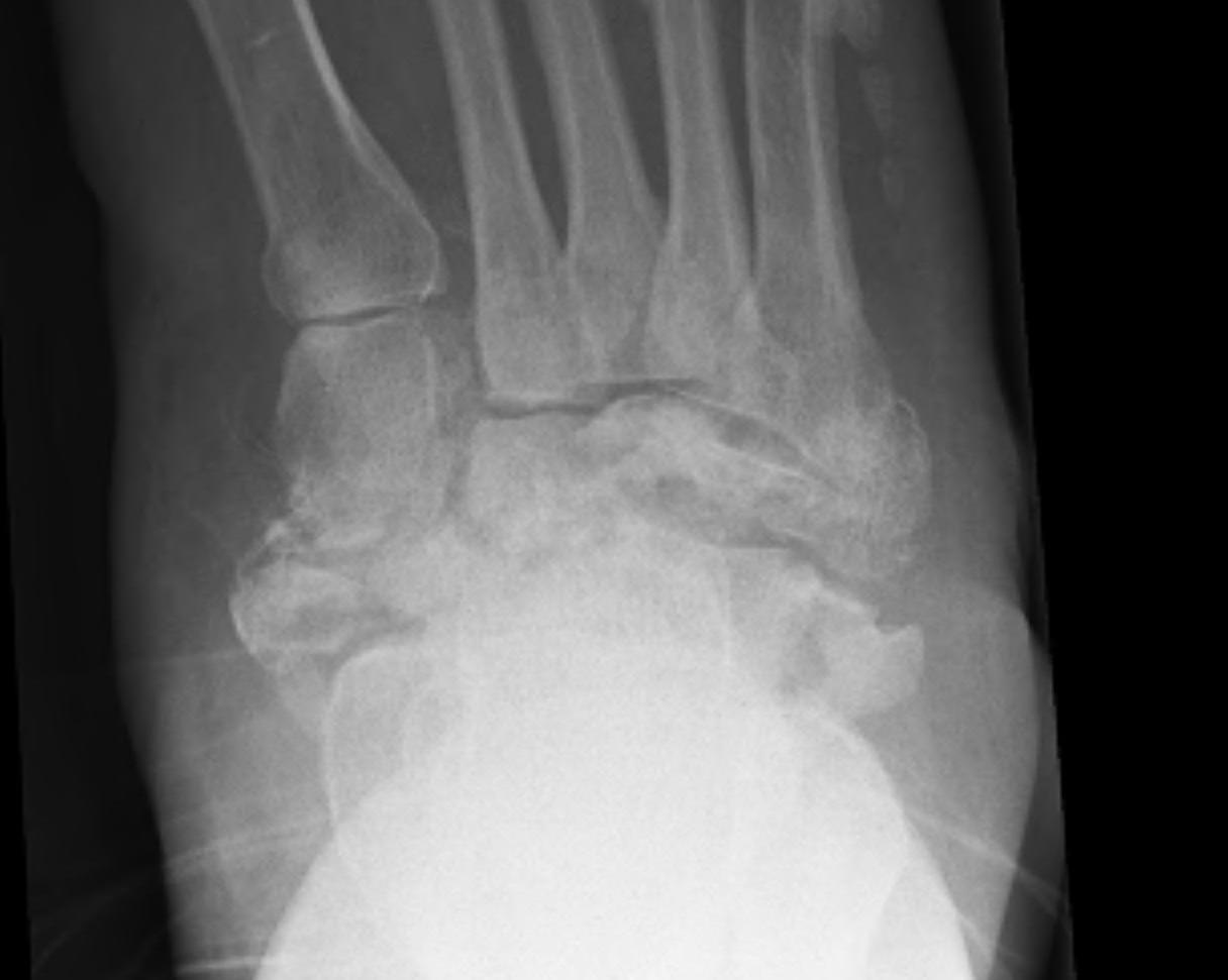
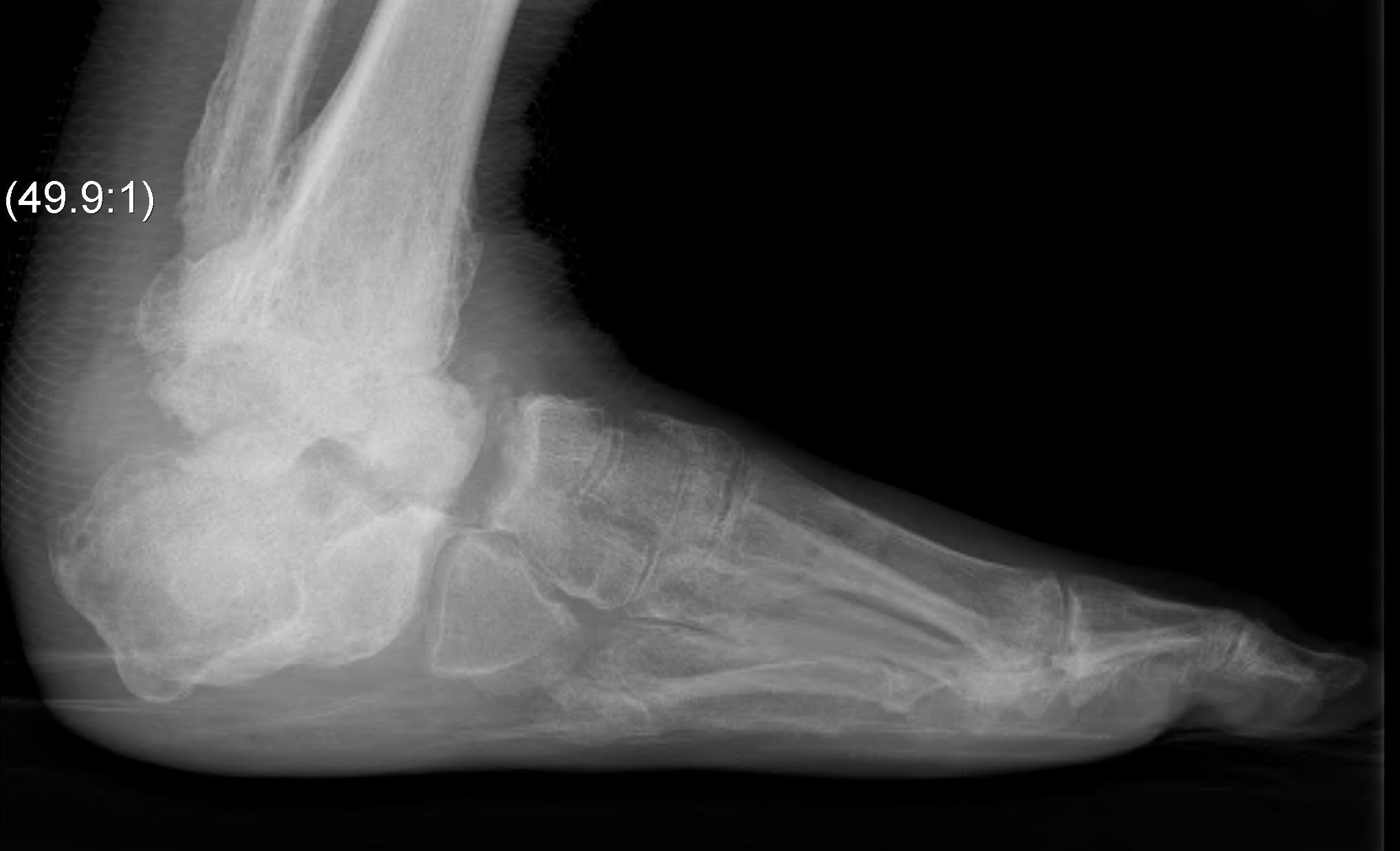
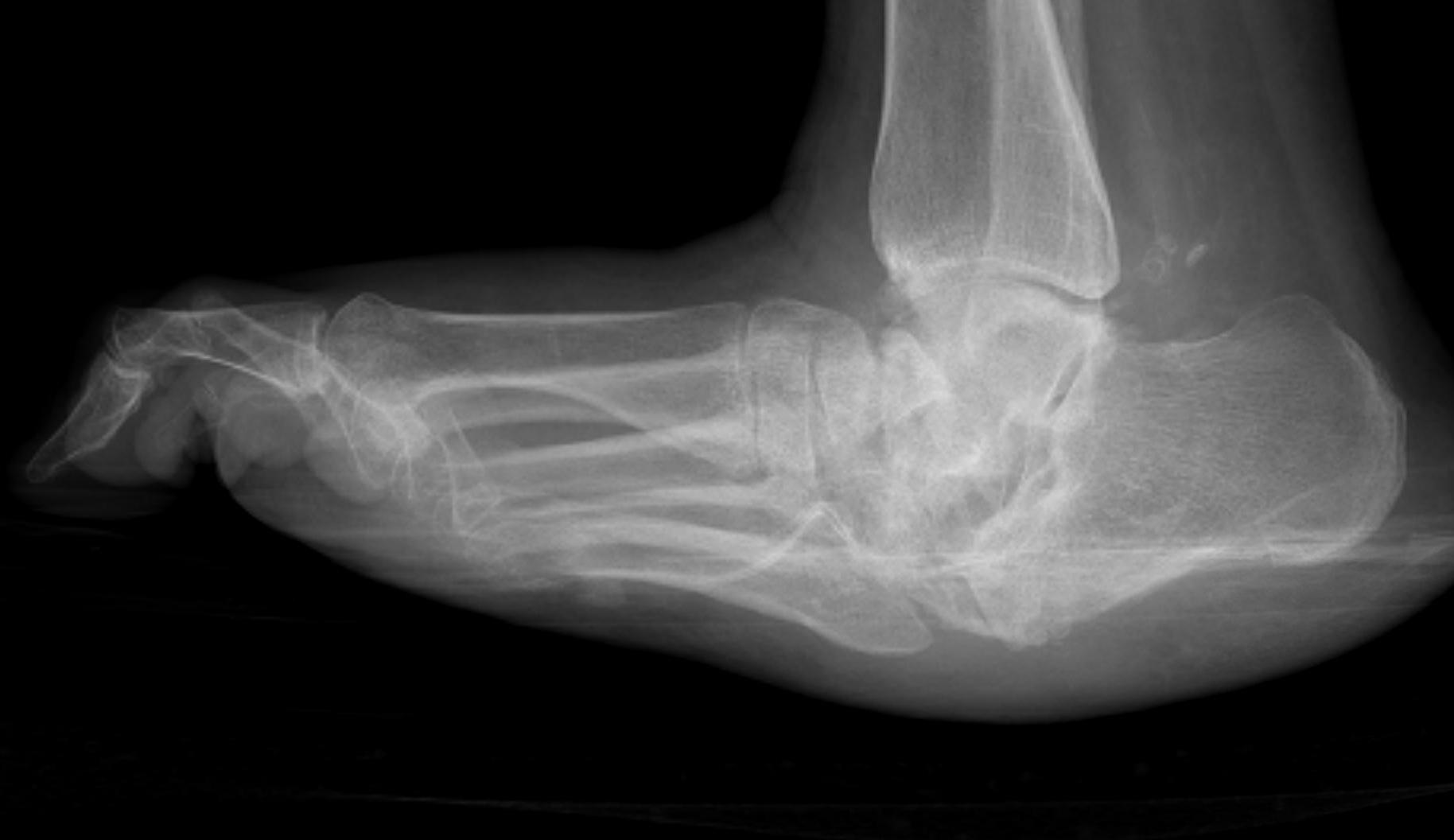
Brodsky Classification
| Type 1 Midfoot (60%) | Type 2 - Hindfoot (30%) | Type 3 (10%) |
|---|---|---|
|
Metatarsocuneiform and naviculocuneiform
Collapse of the medial longitudinal arch with rocker bottom foot |
Subtalar joint, talonavicular, calcaneocuboid
More unstable than type 1 Require longer periods immobilisation |
3a: Tibiotalar joint - most unstable pattern
3b: Fracture calcaneal tubercle - weak push-off and ulceration |
|
 |
Investigation
DDx infection
MRI
Labelled WCC + Bone Scan
Management
Goal
Stable plantigrade foot that is shoe-able or braceable
Few require operative surgery
- control with casts and braces
Indications For Surgery
1. Severe deformity unable to brace
2. Marked instability (usually type II or IIIa)
3. Ulcers
- common type 1
- aim to try and heal ulcer first
- may be caused by fixed bony deformity i.e. midfoot collapse
4. Soft tissues at risk
Contra-Indications
Uncontrolled diabetes
PVD
Medically unwell
Stage 1 disease
Goals of Operative Management
Restore alignment & stability so brace &/or shoe can be worn
- prevent alternative which is amputation
Timing of Surgery
Operating in stage 1 or 2 remains very controversial
Correct deformity in resolution / consolidation stage III
- after cast / brace, shoe failed
Acute Fractures
Issue
- is it charcot or non charcot?
1. Likely Charcot
Patient
- fracture a week or 2 old / red & swollen
- peripheral neuropathy & displaced fracture
- mimimal trauma
Eichenholtz I
- treat non-operatively
2. Non Charcot
Truly acute fracture
- reasonable trauma
- patient has peripheral neuropathy / DM
- treat as per usual, but accept higher complication rate
Management
- ORIF early before acute (dissolution) phase sets in
- if delayed be wary of ORIF as bone stock very poor
- need very strong and augmented ORIF
- must warn of risk of Charcot in acute fracture
- with peripheral neuropathy double period of immobilisation
- NWB 3/12 then further 3-4 month in TCC
Surgical procedures
1. Midfoot ostectomy

Midfoot most common site for neuropathic destruction
- mid foot collapse
- apex of rocker-bottom common site for recurrent ulceration
Technique Ostectomy
1. Attempt to heal ulcer first
- TCC
- debridement +/- IV ABs if OM
2. Remove bony prominence causing ulcer
- medial or lateral incision
- avoid areas of ulceration
- full thickness soft tissue dissection to expose exostosis
- remove with osteotome / saw
- smooth edges with rasp
- haemostasis
- closure over drain; compressive dressing
- postoperative TCC for 6/52
2. Hindfoot Realignment & Arthrodesis
Indications
- hindfoot Charcot not amenable to bracing
- severe deformity or instability following failed bracing
- amputation is only alternative
Amputation v Arthrodesis
May develop bilateral issues
- try to avoid bilateral amputations
Contraindications to Arthrodesis
1. Disease Factors
- Active infection (consider staged)
- Stage I Eichenholtz
- Insufficient soft tissue coverage
- Insufficient bone stock
2. Patient Factors
- Uncontrolled DM or malnutrition
- Nonreconstructable PVD
- Non-compliant
Technique
Preoperative
- cast / TCC till Stage III
- optimise HBA1c and nutrition
Intraoperative
- longitudinal incisions with full thickness flaps under no tension
- meticulous soft tissue handling
- resect bone to correct deformity
- strongest fixation device possible ; often augmented
- if using hindfoot nail ensure >200mm length
(risk of tibial stress fractures with shorter nail)
- often need percutaneous T Achilles lengthening
- alternative: fine wire fixation if active infection
Postoperative
- TCC - 3/12 NWB ; 1/12 PWB; 1/12 WBAT
- Lifelong AFO
- Periodic 6/12 follow-up
Results
- Lowery FAI 2012 - 76% bony fusion; 22% fibrous ; 1.2% amputation
- fibrous union can still result in good function