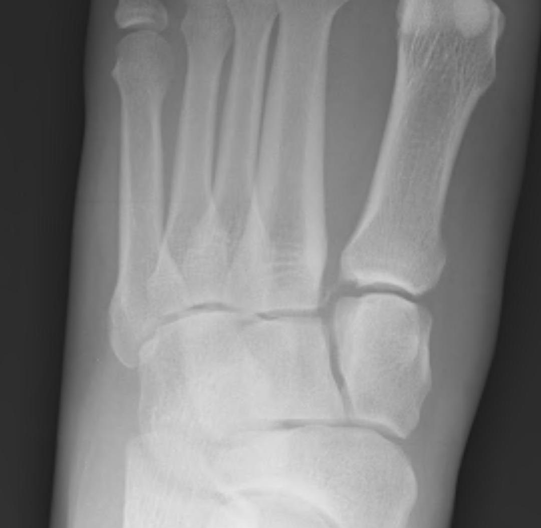


Definition
Injury to the Lisfranc ligament / tarsometarsal joint complex
Low energy injuries - athletes / football / gymnastics
Easily missed
Injury patterns
Lisfranc ligament - medial cuneiform - 2nd metatarsal
Inter-cuneiform instability - medial cuneiform (C1) - middle cuneiform (C2) instabiility
1st TMT joint instability - medial cuneiform (C1) - 1st metatarsal
2nd TMT joint instability - middle cuneiform (C2) - 2nd metatarsal
Porter et al Foot Ankle Int 2019
- 82 athletes with unstable ligamentous Lisfranc injuries
- 50% had inter-cuneiform ligament disruption
Anatomy
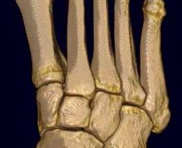
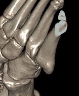
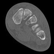
Bony Stability
| Alignment | Alignment | Roman arch | Keystone / Mortise |
|---|---|---|---|
|
1st metatarsal - medial cuneiform 2nd metatarsal - middle cuneiform 3rd metatarsal - lateral cuneiform
AP foot view |
4th and 5th metatarsals articulate with cuboid
Oblique foot view |
Bases of metatarsal wider dorsally than plantar Form half of Roman arch |
2nd metatarsal is keystone of transverse metatarsal arch - middle cuneiform is recessed proximally - mortise provided for base of second metatarsal |
 |
 |
 |
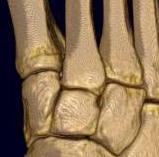 |
Ligamentous stability
| LisFranc Ligament | Interosseous cuneiform ligaments | Tarso-metatarsal joints | Inter-metatarsal ligaments |
|---|---|---|---|
| Plantar, interosseous and dorsal | Plantar, interosseous and dorsal | Plantar and dorsal | Plantar, interosseous and dorsal |
| Base of 2nd metatarsal to medial cuneiform | Medial to intermediate cuneiform | Tarsometatarsal ligamaments | Metatarsal bases |
| Plantar most important | Plantar strongest | Plantar stronger - usually displace dorsally | No connection between 1st and 2nd metatarsal |
Presentation
Significant midfoot pain and swelling
Ecchymosis base of foot over Lisfranc ligament
Imaging
Imaging
1. Diastasis of the intermetatarsal gap between the 1st and 2nd metatarsals
2. Widening of the space between the medial cuneiform and base of 2nd metatarsal
3. Second metatarsal Fleck sign - avulsion of Lisfranc ligament from base of 2nd metatarsal
4. Widening of inter-cuneiform distance
5. Dorsal subluxation of the metatarsals
6. Tarsometatarsal alignment disruption
- medial border 1st metatarsal aligns with medial border medial cuneirform (AP foot)
- medial border 2nd metatarsal aligns with medial border middle cuneiform (AP foot)
- medial border 3rd metatarsal aligns with medial border lateral cuneiform (AP view)
- medial border 4th metatarsal aligns with medial border of the cuboid (oblique view)
X-ray
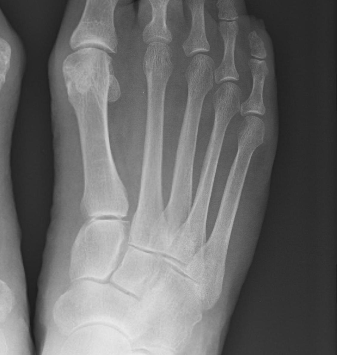
Subtle widening of the medial cuneiform - 2nd metarsal distance, and the inter-metatarsal distance

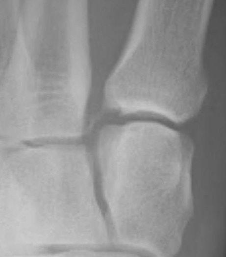
Widening of the medial cuneiform - 2nd metatarsal distance, inter-metatarsal diastasis, fleck sign, possibly increased inter-cuneiform distance
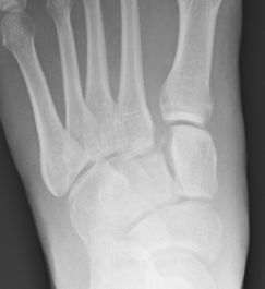
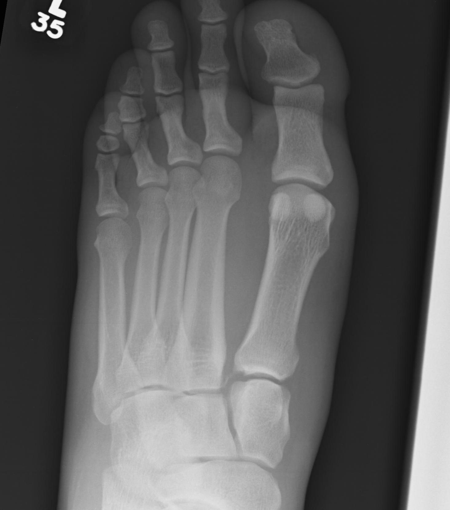
Widening of the medial cuneiform - 2nd metatarsal distance, inter-metatarsal diastasis, fleck sign, increased inter-cuneiform distance
Weight bearing xrays
Sripanich et al Skeletal Radiol 2020
- systematic review
- weight bearing xrays more sensitive at detecting subtle Lisfranc instability


Increased widening of inter-metatarsal distance on weight bearing views


Increased widening of Lisfranc joint with subluxation of the 1st and 2nd metatarsals on weight bearing views
CT


Fleck sign


Fleck sign with inter-cuneiform widening
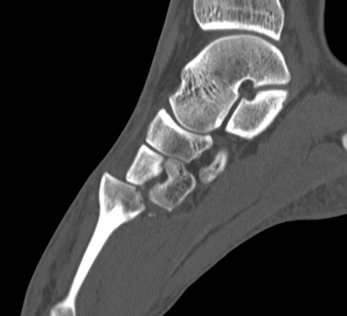
Dorsal subluxation of the metatarsal
Weight bearing CT
Talaski et al Foot Ankle Orthop 2023
- systematic review of CT versus weight bearing CT
- weight bearing CT more sensitive at detecting subtle Lisfranc instability
MRI
Diagnose
- Lisfranc ligament injury
- inter-cuneiform disruption
- 31 subtle Lisfranc confirmed at surgery
- correct diagnosis: xray 48%, CT 87%, MRi 97%
Management
Non operative Management
- systematic review of nonoperative management of Lisfranc injuries
- heterogenous group
- superior results with surgery
- late displacement up to 54% with nonoperative management
- surgical intervention up to 56% with nonoperative management
- best outcome of nonoperative management with no displacement on CT
Operative Management
Indications
Any displacement
Any instability with weight bearing
Options
Trans-articular screw fixation
Flexible fixation - suture button / internal brace
Bridge plate fixation
Primary Arthrodesis


Results
Screw versus suture button
- 60 isolated Lisfranc ligament injuries
- half treated with screw, half with suture button
- no difference in functional outcome at 1 year
- recurrent diastasis with 1/30 screws and 2/30 suture buttons
Screw versus suture button versus primary arthrodesis
- systematic review and meta-analysis isolated Lisfranc ligament injuries
- flexible fixation versus ORIF versus primary arthrodesis
- 25 studies and 300 patients
- better outcomes and return to activity with flexible fixation
Screw versus primary arthrodesis
- RCT of 40 patients with ligamentous Lis Franc injuries
- better outcomes with primary arthrodesis
Screw versus bridge plating
- comparison of screw fixation versus bridge plating
- 60 ligamentous Lis Franc
- slightly better outcome scores at 2 years with bridge plate
- anatomic reduction: bridge plate 91%, screw 82%
Screw versus primary arthrodesis 1st TMT dislocation
- 78 patients with 1st TMT dislocation and Lisfranc
- ORIF versus primary arthrodesis
- improved functional outcomes with arthrodesis
- loss of reduction in 25% of ORIF
Bridge plate versus primary arthrodesis 1st TMT dislocation
Stodle et al Foot Ankle Int 2020
- Lisfranc with 1st tarsometatarsal dislocation
- RCT of temporary plate fixation versus arthrodesis
- 48 patients
- no difference in outcome at 2 years
Approach and reduction
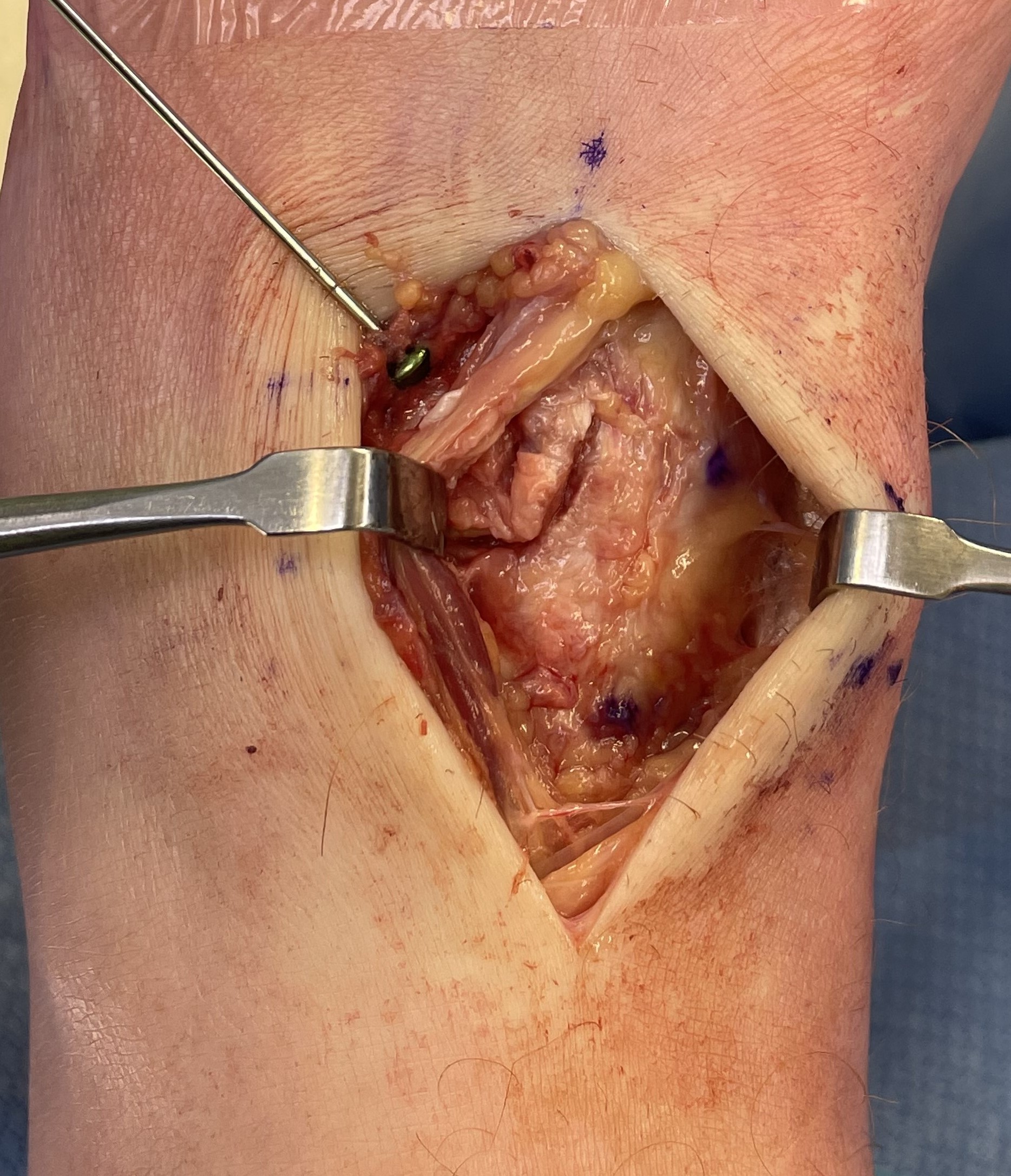
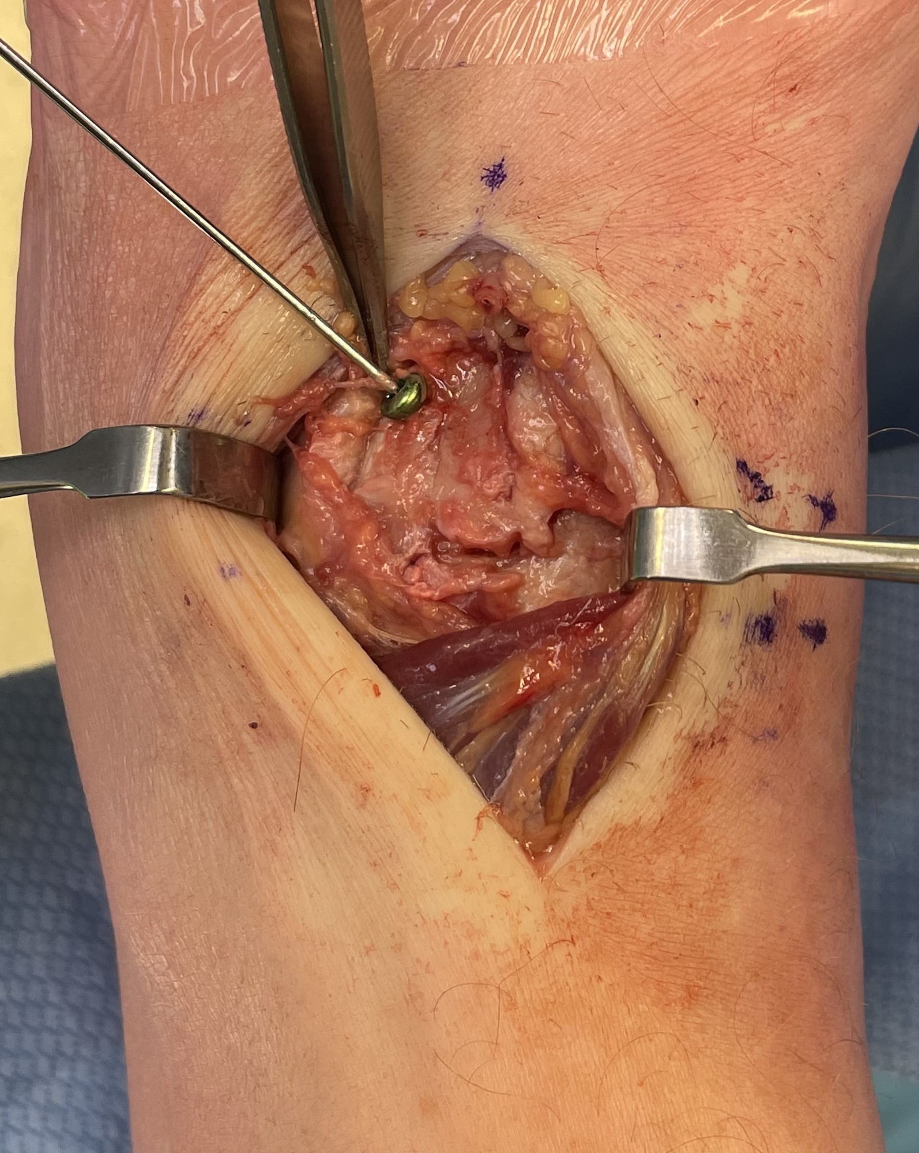
Dorsal incision between 1st and 2nd metatarsal
- protect branches of superficial peroneal nerve
- retract EHL medially
- window either side of EDB / dorsalis pedis and deep peroneal nerve
- retract EDB either way to approach 1st TMT / 2nd TMT
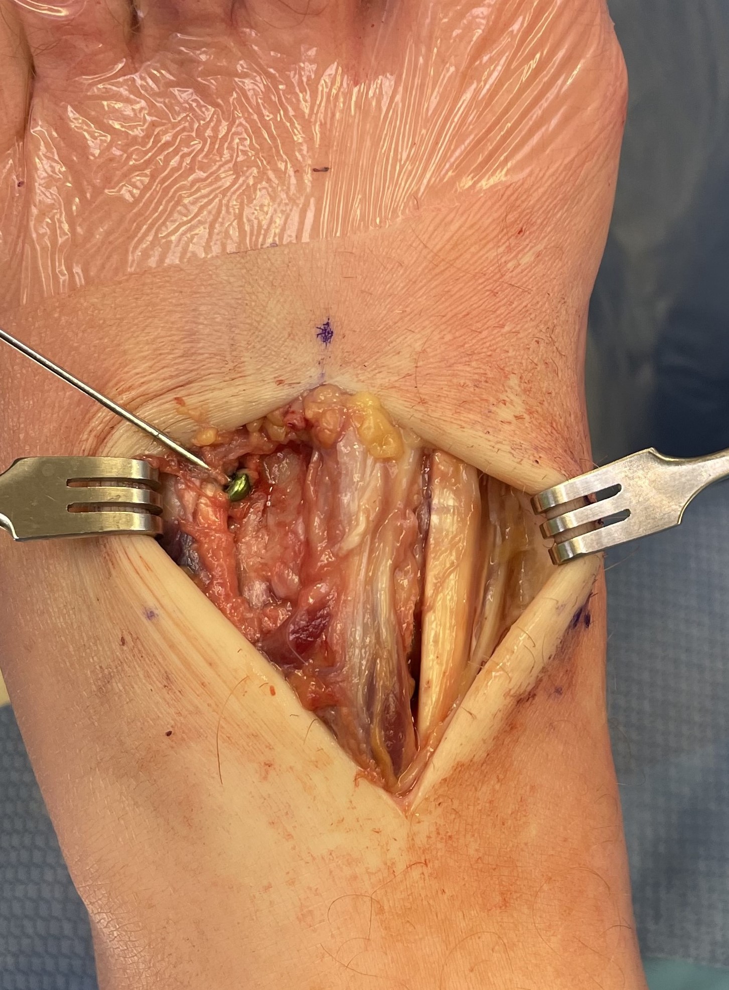
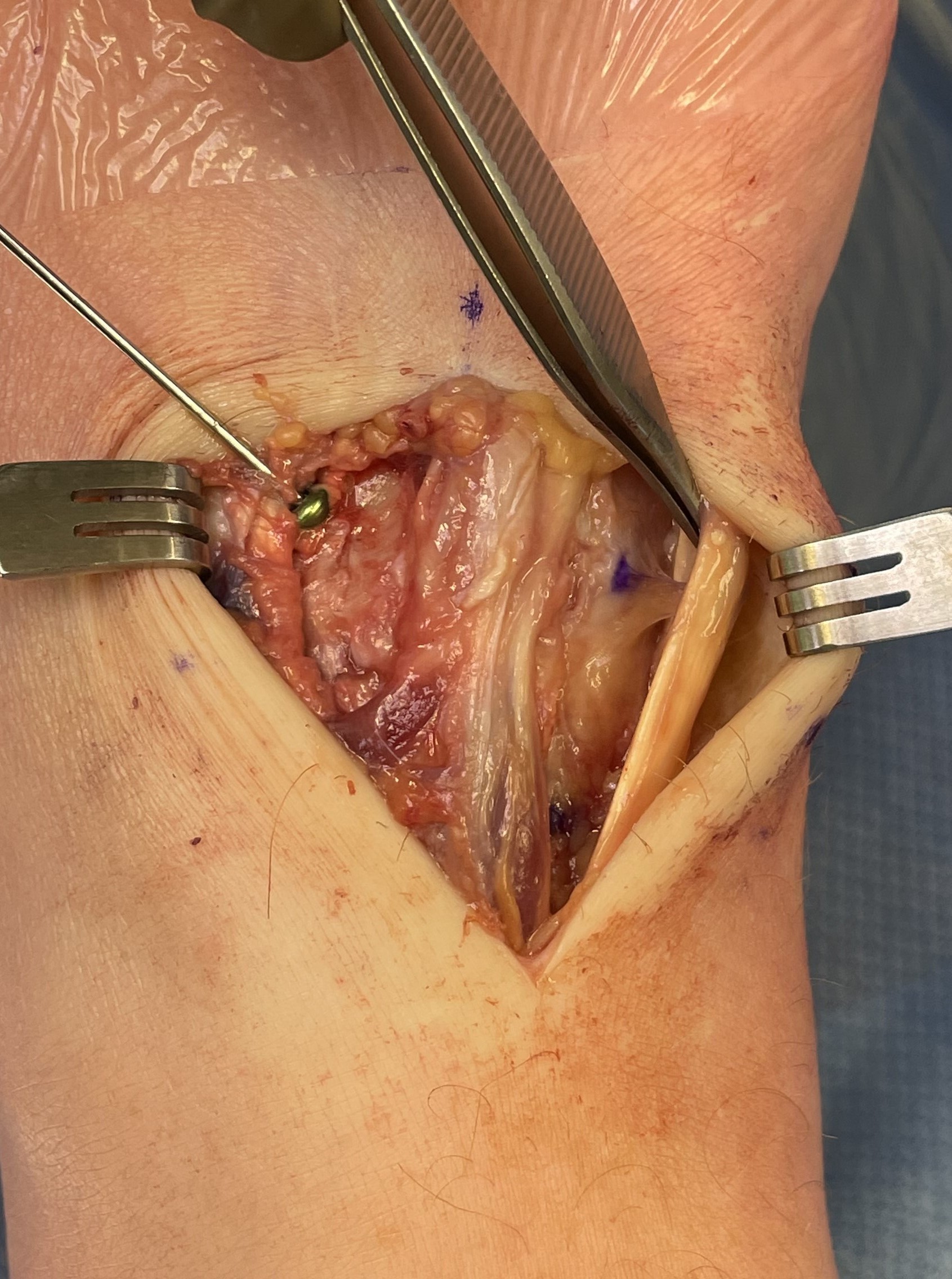
Reduction
- medial incision over medial cuneiform
- clamp medial cuneiform to base 2nd metatarsal
Screw fixation
AP view
- 1st metatarsal to medial cuneiform - screw
- 2nd metatarsal to intermediate cuneiform - screw
- medial cuneiform to base of second metatarsal - screw
+/- medial cuneiform to intermediate cuneiform - screw


Screw removal
| Pros | Cons |
|---|---|
|
Second surgery Infection / nerve injury Loss of reduction |
Broken screws Increased difficulty with any subsequent surgery |
Rhodes et al Foot Ankle Orthop 2022
- systematic review of routine metalwork removal versus retention in Lisfranc surgery
- 28 studies and 1000 patients
- marginal improvement in function with routine removal


Suture button fixation


Arthrex mini tightrope Lisfranc PDF
Arthrex mini tightrope Lisfranc video
Internal Brace


Arthrex internal brace LisFranc PDF
Arthrex internal brace LisFranc video
Vumedi Flexible Fixation Lisfranc injury video
Bridge plate
Advantage
- avoid articular cartilage damage
- avoid broken screws across joint


Primary Arthrodesis


Missed Lisfranc
Options
Ligamentous reconstruction
Arthrodesis
